

Fundus photography eye test involves capturing a photograph of the back of the eye, i.e. fundus or the retina, all the retina blood vessels, and the optic nerve. The fundus eye test is frequently used to document the retina condition, and is also called the colour fundus photograph and forms an essential step in treating diseases of the retina.
A fundus eye test and retina pictures are not standalone investigations. Fundus photography is one of the ways to perform retinal imaging. A few others, too, like OCT and Fundus fluorescein angiography.
At the outset, the patient has to undergo a general eye examination. Here instruments like the slit lamp and applanation tonometer are used. After dilating the pupil, your eye doctor or retina specialist also uses an indirect ophthalmoscope to examine your retina. The retina is also known as the fundus. Once the fundus examination is done, a decision is taken when the fundus picture is needed or not.
Fundus cameras that consist of a microscope attached to a flash-enabled camera are used in fundus eye tests. The main structures visualized on a fundus photo are the central and peripheral retina, optic disc or optic nerve head, and macula or posterior pole. Fundus photography can be performed with coloured filters or specialized dyes, including fluorescein and indocyanine green.
Fundus photography is done after a fundus examination. It is used to inspect anomalies that affect the eyes and blood vessels of the retina and monitor these diseases' progression. It is vital for disease processes such as macular degeneration, retinal neoplasms, choroid disturbances and diabetic eye disease, and associated macular oedema. Additionally, it aids in identifying glaucoma, multiple sclerosis, and other central nervous system abnormalities.
It evaluates irregularities in the fundus and monitors the progression of a disease. This fundus eye test also informs both patients and eye doctors about the management and treatment outcome. They are crucial to create a starting point or baseline to understand a disease's progression better. Fundus photographs may be helpful if there is a new disease affecting the fundus and planning additional treatment options.
Your eye doctor or retina specialist must record these photos and others in an orderly fashion so that they can compare photographs of a patient from different timelines.
FFA or Fundus Fluorescein Angiography is a procedure that comprises injecting a dye, FLUORESCEIN, into one of your veins in the arm. After this injection, we take fundus photos of the dye's passage within the inner structures of the eye – the retina and choroid.
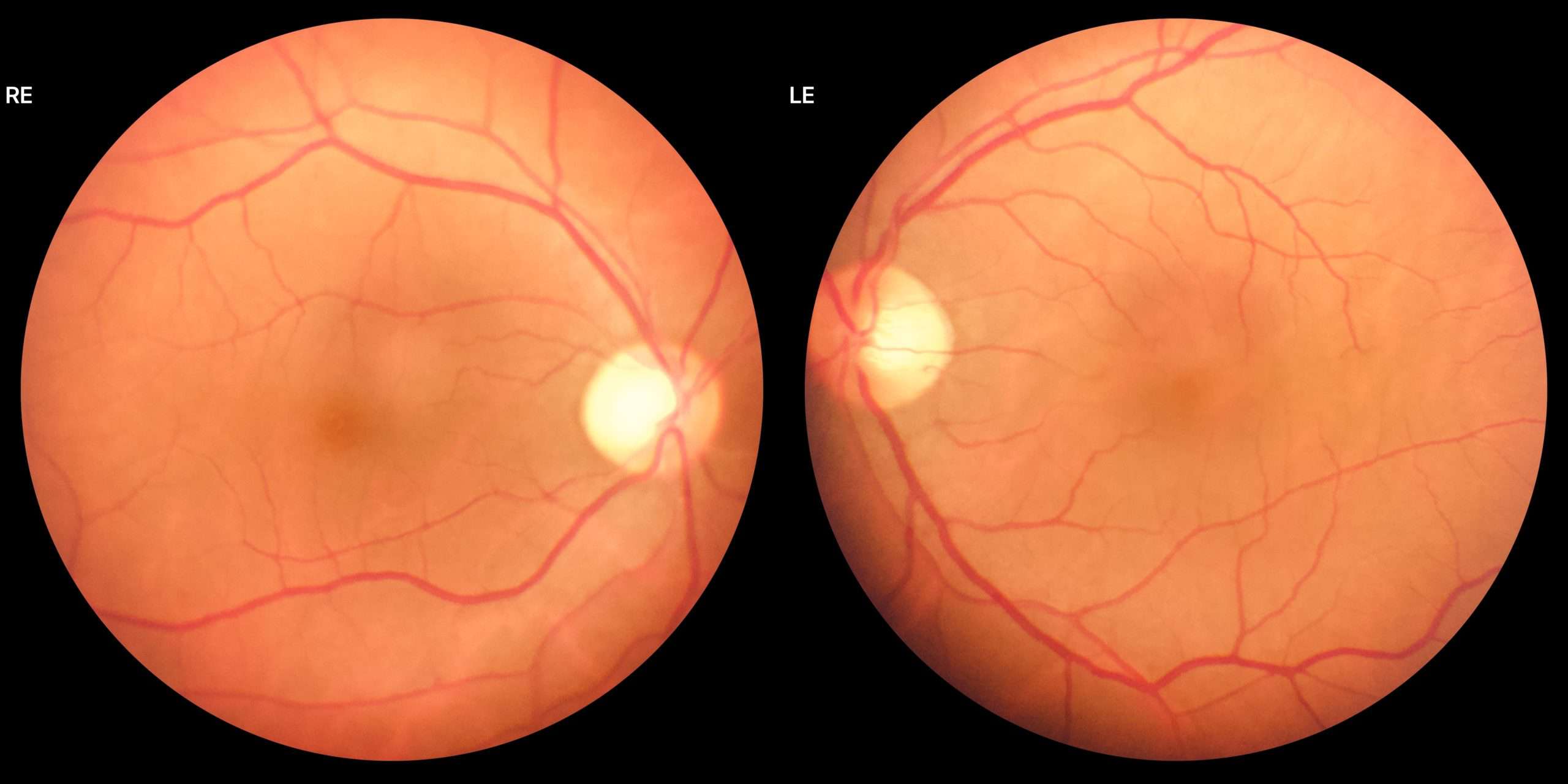
After a routine dilated eye examination, the doctor may advise you to get this test done. There are many kinds of fundus cameras available in the market. The one we have fits on the slit lamp itself, so the patient does not have to get up from their seat. After bringing the camera into position, the patient positions their head such that the chin rests on the chin rest and the forehead is against the headband. The camera is focused on the retina, and as you can see in the video below, the picture of the retina is captured. While focussing, the camera will come very close to the eye's surface but not touch the eye. From the picture below, you can see the optic nerve, the retina, and the retinal blood vessels. The image that you see above is that of a normal-looking retina.
As mentioned earlier, this picture is immediately attached to the patient's records to view anytime in the future.
